
Mouse Secondary Antibodies

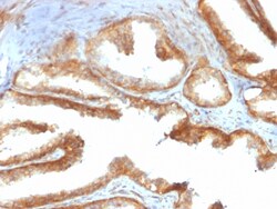
Biotium YBX1 / Y-box Binding Protein 1 / YB-1 (Tumor Biomarker)(rYBX1/2430), 1mg/mL
The specificity of this monoclonal antibody to its intended target was validated by HuProt™ Array, containing more than 19,000 full-length human proteins. Y-box binding protein-1 (YBX1) is the prototypic member of the cold shock protein family that fulfills numerous cellular functions. In the nucleus, YBX1 protein orchestrates transcription of proliferation-related genes, whereas in the cytoplasm it associates with mRNA and directs translation. In human tumor entities, such as breast, lung and prostate cancer, cellular YBX1 expression indicates poor clinical outcome, suggesting that YBX1 is an attractive marker to predict patients' prognosis. Primary antibodies are available purified, or with a selection of fluorescent CF Dyes and other labels. CF Dyes offer exceptional brightness and photostability. Note: Conjugates of blue fluorescent dyes like CF405S and CF405M are not recommended for detecting low abundance targets, because blue dyes have lower fluorescence an

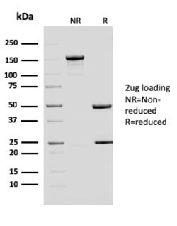
Biotium ERCC1 / RAD10 (Tumor Progression Marker)(ERCC1/2683), CF488A conjugate, 0.1mg/mL
Recognizes a protein of 110 kDa, identified as Excision Repair Cross Complementing 1 (ERCC1). It is a mammalian nucleotide excision repair (NER) enzyme involved in repair of damaged DNA. ERCC1 is a homologous to RAD10 in Saccharomyces cerevisiae, which is required in mitotic intrachromosomal recombination and repair. ERCC1 is required in repair of cisplatin-induced DNA adducts and ultraviolet (UV)-induced DNA damage. High expression of ERCC1 has been linked to tumor progression in a variety of cancers including non-small cell lung cancer (NSCLC), squamous cell carcinoma of the head, ovarian cancer and esophageal cancer. Primary antibodies are available purified, or with a selection of fluorescent CF Dyes and other labels. CF Dyes offer exceptional brightness and photostability. Note: Conjugates of blue fluorescent dyes like CF405S and CF405M are not recommended for detecting low abundance targets, because blue dyes have lower fluorescence and can give higher non-specific

Biotium Gastrin(GAST/2631), 0.2mg/mL
Gastrin, is a hormone that normally formed by mucosal cells in the gastric antrum and by the D cells of the pancreatic islets. Its primary function is to stimulate secretion of HCl by the gastric mucosa. HCl, in turn, inhibits gastrin formation. It also responsible for stimulating smooth muscle contraction and increasing blood circulation and water secretion in the stomach and intestine. Gastrin is regulated by epidermal growth factor in both mice and humans. Gastrin is excreted in excess by pancreatic tumors in the Zollinger-Ellison syndrome. Gastrin-Releasing Peptide (GRP) stimulates the release of gastrin as well as other gastrointestinal hormones and also acts as an autocrine growth factor for certain cell types. High levels of GRP are found in the human lung just after birth and levels decrease thereafter in parallel with the observed disease in a number of pulmonary neuroendocrine cells. GRP is known to promote lung tumorigenesis in model systems. Primary antibodies are
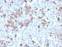
Biotium Prolactin (Pituitary Tumor Marker)(PRL/2642), Biotin conjugate, 0.1mg/mL
Prolactin is a growth factor that is secreted by the anterior pituitary. It is necessary for the proliferation and differentiation of the mammary glands. Prolactin is useful in the classification of pituitary tumors and study of pituitary disease. It also plays a role in the development of mammary cancer, functioning dually as a mitogen and a differentiating agent. Primary antibodies are available purified, or with a selection of fluorescent CF Dyes and other labels. CF Dyes offer exceptional brightness and photostability. Note: Conjugates of blue fluorescent dyes like CF405S and CF405M are not recommended for detecting low abundance targets, because blue dyes have lower fluorescence and can give higher non-specific background than other dye colors.
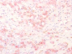
Biotium Tyrosinase-Related Protein-1 (TYRP-1) (Melanoma Marker), Biotin conjugate, 0.1mg/mL
Reacts with a 75 kDa melanocyte-specific gene product, identified as Tyrosinase-related protein-1 (TRP-1). It is involved in melanin synthesis. TRP1 is present on the melanosomal membranes of melanoma, normal melanocytes and nevi. Recent evidence suggests that TRP-1 is involved in maintaining stability of tyrosinase protein and modulating its catalytic activity. TRP-1 is also involved in maintenance of melanosome ultrastructure and affects melanocyte proliferation and cell death. Primary antibodies are available purified, or with a selection of fluorescent CF Dyes and other labels. CF Dyes offer exceptional brightness and photostability. Note: Conjugates of blue fluorescent dyes like CF405S and CF405M are not recommended for detecting low abundance targets, because blue dyes have lower fluorescence and can give higher non-specific background than other dye colors.
Biotium Peroxiredoxin 4 (Prognostic Marker for Lung SqCC)(CPTC-PRDX4-2), CF488A conjugate, 0.1mg/mL
The peroxiredoxin (PRX) family comprises six antioxidant proteins, PRX I, II, III, IV, V and VI, which protect cells from reactive oxygen species (ROS) by preventing the metal-catalyzed oxidation of enzymes. The PRX proteins primarily utilize thioredoxin as the electron donor for antioxidation, although they are fairly promiscuous with regard to the hydroperoxide substrate. In addition to protection from ROS, peroxiredoxins are also involved in cell proliferation, differentiation and gene expression. PRX I, II, IV and VI show diffuse cytoplasmic localization, while PRX III and V exhibit distinct mitochondrial localization. The human PRX IV gene is expressed in many tissues. It exists as a precursor protein, which is only detected in testis, and aprocessed secreted form. PRX IV is highly expressed in lung cancer and is necessary for the promotion of lung cancer in vitro. Studies have demonstrated that PRX IV positive expression is significantly correlated with recurrences and
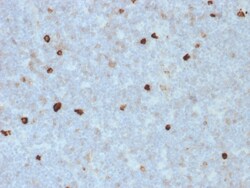
Biotium Human IgM (Immunoglobulin Mu Heavy Chain) (B-Cell Marker)(R1/69), CF405S conjugate, 0.1mg/mL
Recognizes a protein of 75 kDa, identified as mu heavy chain of human immunoglobulins. It does not cross-react with alpha (IgA), gamma (IgG), epsilon (IgE), or delta (IgD), heavy chains, T-cells, monocytes, granulocytes, or erythrocytes. Monomeric IgM is expressed as a membrane bound antibody on the surface of B cells and as a pentamer when secreted by plasma cells. IgM antibody is prominent in early immune responses to most antigens. Aberrant levels are associated with immune deficiency states, hereditary deficiencies, myeloma, Waldenstrom's macroglobulinemia, chronic infection and hepatocellular disease. This MAb is useful in the identification of leukemias, plasmacytomas, and certain non-Hodgkin's lymphomas. The most common feature of these malignancies is the restricted expression of a single heavy chain class. Demonstration of clonality in lymphoid infiltrates indicates that the infiltrate is clonal and therefore malignant. Primary antibodies are available purified, or with a
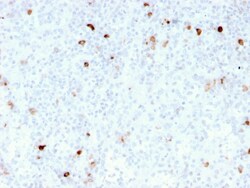
Biotium IgM (Immunoglobulin Mu Heavy Chain) (B-Cell Marker)(rIM260), CF740 conjugate, 0.1mg/mL
Recognizes a protein of 75 kDa, identified as mu heavy chain of human immunoglobulins. It does not cross-react with alpha (IgA), gamma (IgG), epsilon (IgE), or delta (IgD), heavy chains, T-cells, monocytes, granulocytes, or erythrocytes. This MAb is useful in the identification of leukemias, plasmacytomas, and certain non-Hodgkin's lymphomas. The most common feature of these malignancies is the restricted expression of a single heavy chain class. Demonstration of clonality in lymphoid infiltrates indicates that the infiltrate is clonal and therefore malignant. Primary antibodies are available purified, or with a selection of fluorescent CF Dyes and other labels. CF Dyes offer exceptional brightness and photostability. Note: Conjugates of blue fluorescent dyes like CF405S and CF405M are not recommended for detecting low abundance targets, because blue dyes have lower fluorescence and can give higher non-specific background than other dye colors.
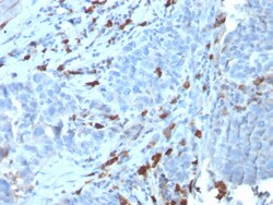
Biotium Human IgM (Immunoglobulin Mu Heavy Chain) (B-Cell Marker)(IGHM/3135R), CF647 conjugate, 0.1mg/mL
Recognizes a protein of 75 kDa, identified as mu heavy chain of human immunoglobulins. It does not cross-react with alpha (IgA), gamma (IgG), epsilon (IgE), or delta (IgD), heavy chains, T-cells, monocytes, granulocytes, or erythrocytes. Monomeric IgM is expressed as a membrane bound antibody on the surface of B cells and as a pentamer when secreted by plasma cells. IgM antibody is prominent in early immune responses to most antigens. Aberrant levels are associated with immune deficiency states, hereditary deficiencies, myeloma, Waldenstrom's macroglobulinemia, chronic infection and hepatocellular disease. This MAb is useful in the identification of leukemias, plasmacytomas, and certain non-Hodgkin's lymphomas. The most common feature of these malignancies is the restricted expression of a single heavy chain class. Demonstration of clonality in lymphoid infiltrates indicates that the infiltrate is clonal and therefore malignant. Primary antibodies are available purified, or w
Miltenyi Biotec, Inc. CD86-APC, mouse, REA1190, 100 tests
Identification and enumeration of CD86+ cells by flow cytometry.
This product is subject to the following: Miltenyi Biotec, Inc. Terms and Conditions of Sale For Products and Services
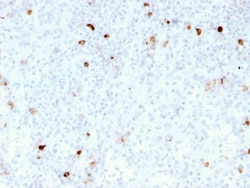
Biotium IgM (Immunoglobulin Mu Heavy Chain) (B-Cell Marker)(rIM260), CF405S conjugate, 0.1mg/mL
Recognizes a protein of 75 kDa, identified as mu heavy chain of human immunoglobulins. It does not cross-react with alpha (IgA), gamma (IgG), epsilon (IgE), or delta (IgD), heavy chains, T-cells, monocytes, granulocytes, or erythrocytes. This MAb is useful in the identification of leukemias, plasmacytomas, and certain non-Hodgkin's lymphomas. The most common feature of these malignancies is the restricted expression of a single heavy chain class. Demonstration of clonality in lymphoid infiltrates indicates that the infiltrate is clonal and therefore malignant. Primary antibodies are available purified, or with a selection of fluorescent CF Dyes and other labels. CF Dyes offer exceptional brightness and photostability. Note: Conjugates of blue fluorescent dyes like CF405S and CF405M are not recommended for detecting low abundance targets, because blue dyes have lower fluorescence and can give higher non-specific background than other dye colors.
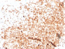
Biotium Human IgM (Immunoglobulin Mu Heavy Chain) (B-Cell Marker)(rIGHM/3802), CF740 conjugate, 0.1mg/mL
Recognizes a protein of 75 kDa, identified as mu heavy chain of human immunoglobulins. It does not cross-react with alpha (IgA), gamma (IgG), epsilon (IgE), or delta (IgD), heavy chains, T-cells, monocytes, granulocytes, or erythrocytes. Monomeric IgM is expressed as a membrane bound antibody on the surface of B cells and as a pentamer when secreted by plasma cells. IgM antibody is prominent in early immune responses to most antigens. Aberrant levels are associated with immune deficiency states, hereditary deficiencies, myeloma, Waldenstrom's macroglobulinemia, chronic infection and hepatocellular disease. This MAb is useful in the identification of leukemias, plasmacytomas, and certain non-Hodgkin's lymphomas. The most common feature of these malignancies is the restricted expression of a single heavy chain class. Demonstration of clonality in lymphoid infiltrates indicates that the infiltrate is clonal and therefore malignant. Primary antibodies are available purified, or w

Biotium Human IgM (Immunoglobulin Mu Heavy Chain) (B-Cell Marker)(rIGHM/1623), 0.2mg/mL
Recognizes a protein of 75 kDa, identified as mu heavy chain of human immunoglobulins. It does not cross-react with alpha (IgA), gamma (IgG), epsilon (IgE), or delta (IgD), heavy chains, T-cells, monocytes, granulocytes, or erythrocytes. Monomeric IgM is expressed as a membrane bound antibody on the surface of B cells and as a pentamer when secreted by plasma cells. IgM antibody is prominent in early immune responses to most antigens. Aberrant levels are associated with immune deficiency states, hereditary deficiencies, myeloma, Waldenstrom's macroglobulinemia, chronic infection and hepatocellular disease. This MAb is useful in the identification of leukemias, plasmacytomas, and certain non-Hodgkin's lymphomas. The most common feature of these malignancies is the restricted expression of a single heavy chain class. Demonstration of clonality in lymphoid infiltrates indicates that the infiltrate is clonal and therefore malignant. Primary antibodies are available purified, or w

Cayman Chemical Acylated Ghrelin mouse rat
This EIA kit specifically measures the acylated form of ghrelin.
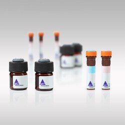
Miltenyi Biotec, Inc. Mouse IgG2b-Biotin, IS6-11E5.11, 30 t
Isotype control antibody for flow cytometric analysis of cells.
This product is subject to the following: Miltenyi Biotec, Inc. Terms and Conditions of Sale For Products and Services